
Self-management through a positive lifestyle It is not advisable to attempt radiosurgery on those with hereditary disease due to the risk of developing additional lesions following radiation. According to the CCM clinical care guidelines, stereotactic radiosurgery should only be considered for solitary lesions that have previously hemorrhaged and carry a high surgical risk. Radiosurgery is a non-surgical targeted treatment for CCM. Surgical resection may be considered if the lesion is symptomatic and has bled on at least one occasion. Surgery should be performed by a surgeon with CCM and skull-base surgery expertise. Surgical removal should be considered on a case-by-case basis with the risks and benefits weighed. As long as the lesion appears stable and there are no additional symptoms or evidence of hemorrhage, this is usually the most prudent course of action.

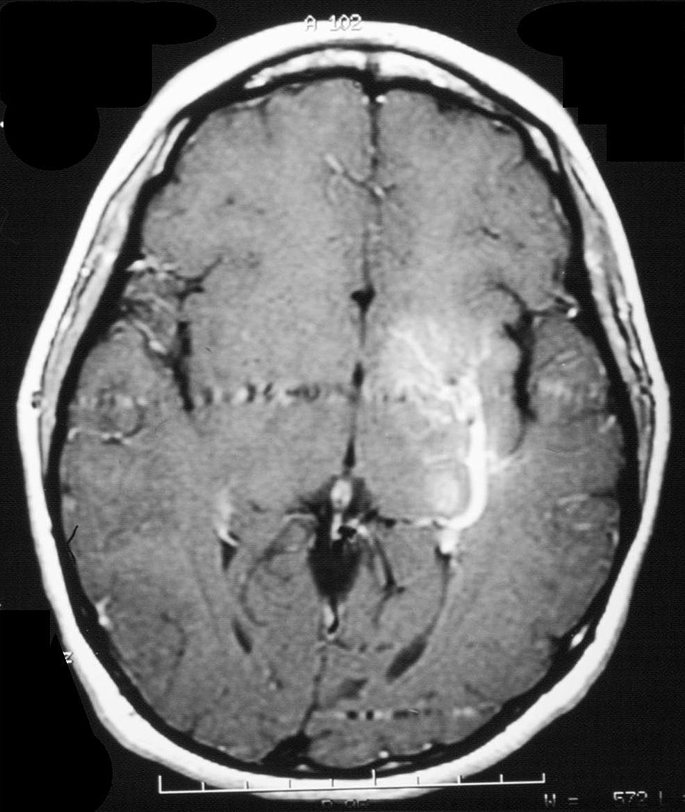
Conservative management consists of routine, periodic MRIs to monitor the changes in the lesion. This approach is most often recommended, especially in lesions that have not bled or are asymptomatic. This is a great resource to share with your providers so that you can make the best decisions regarding your care. The current recommendations for the management of brainstem lesions are outlined in the Clinical Care Guidelines. Management of brainstem cavernous malformation Breathing difficulties that can contribute to sleep issues, including sleep apnea.Hiccups as a result of diaphragmatic spasms.Coordination and movement challenges, or ataxia.These focal neurological deficits can include: The most common symptoms for brainstem lesions are focal neurological deficits, as opposed to seizure or headache for lesions located in other regions of the brain. Other studies show varying rates of re-bleeding. A study measured re-hemorrhaging rates as high as 30% per person.In this study, brainstem hemorrhage location was most common in patients who presented with hemorrhage (58%). A paper published in 2020 (Flemming et al) found that brainstem location is a strong predictor of hemorrhage. Studies have indicated that brainstem lesions are more likely to present with an initial hemorrhage and are more likely to rebleed.Approximately 20% of cavernous malformations are located in the brainstem.Damage to this area can cause more serious effects than other locations within the brain and is associated with greater morbidity and mortality. All rights reserved.The brainstem is a small region packed with nerve fibers that control voluntary and involuntary functions in the body, including regulation of heart rate, breathing, sleeping, and temperature. Surgical or endovascular obliteration of DVAs carries a significant risk of venous infarction thus, conservative management is the treatment of choice for patients with these lesions.ĭevelopmental venous anomaly venous angioma venous malformation venous medullary malformation. Although the natural history of DVAs is benign, they may be associated with cavernous malformations or other vascular abnormalities, which can lead to hemorrhage in the vicinity of the DVA. DVAs are congenital lesions thought to arise from aberrations that occur during venous development, but continue to provide the normal venous drainage to the cerebral territory in which they reside. They are the most common type of cerebral vascular malformation found on autopsy studies, and they are often encountered as incidental findings on neuroimaging studies. DVAs are also known as cerebral venous angiomas, cerebral venous malformations, and cerebral venous medullary malformations. The defining characteristic of these lesions is the confluence of radially oriented veins into a single dilated venous channel. Developmental venous anomalies (DVAs) are relatively common lesions, present in up to 3% of the population.


 0 kommentar(er)
